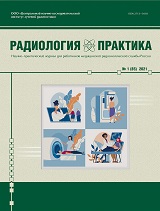
The journal «Radiology — practice» is being published since 2000. The main goal of the issue is coverage of modern technologies and the equipment which aims radiologic images analyses, methods of clinical application: radiography, MRI, CT, ultrasound and radionuclide investigations. We make a scope of continuing education and preparation of x-ray specialists, standardization of all kinds modern x-ray examinations, objective accreditation of x-ray diagnostic departments, and certification, licensing and specialists attesting.
We give medical-technical reviews, such as equipment, examinations methodology, radiation safety, and labour protection. The Journal is intended for x-ray doctors, engineers, medical assistants, technical personnel, dosimetricians, all the leading specialists in x-ray diagnosis, departments’ chiefs in this sphere, chief doctors, and leaders of city/republic level who develop equipment policy in healthcare system.
Target audience: radiologists, specialists of ultrasonic and radionuclide diagnostics, scientists and teachers of specialized departments of universities.
Уважаемые авторы!
Журнал Радиология – практика прекращает принимать на рассмотрение и публикацию обзорные статьи.
Имеющиеся в редакции обзорные работы, прошедшие рецензирование и имеющие положительные отзывы, будут опубликованы в 5-м и 6-м номерах. Оба номера уже сформированы.
Продолжается прием оригинальных статей и клинических наблюдений, которые при наличии положительных отзывов рецензентов будут опубликованы в 2027 году.
С уважением, редакция журнала Радиология – практика.
Current issue
ORIGINAL RESEARCH
Objective. Study of the Density Characteristics of the Cartilaginous Part of the Nasal Septum in Normal, with its Perforation and Deviation using Multispiral Computed Tomography.
Materials and Methods. Multispiral Computed Tomography (MSCT) of the Nasal Septum (NS) and Sinuses was used in the Examination of 125 patients (61 women and 64 men aged 18 to 67 years). Of these, 57 patients (23 women and 34 men) with nasal septum deformity (NSD), 48 patients (28 women and 20 men) with nasal septum perforation (NSP) were performed. The control group included 20 subjects (10 women and 10 men) without NS pathology. MSCT scans were performed on a Philips Ingenuity 64 multispiral tomography scanner. The Protocol included the study of NSD variants, localization and size of NSP, nasal septum density in HU units. In cases of NSP, the density of the cartilaginous region was studied around the perforation. All statistical analyses were performed using the SPSS 23.0 software package.
Results. The data obtained on NSD variants: 1. C-shaped deviation of 9 cases (15.8 % of all cases); 2. S-shaped curvature — 21 cases (36.8 %); 3. «Crest» — 11 cases (19.3 %); 4. Dislocation of the quadrangular cartilage — 2 cases (3.5 %); 5. Thickening — 14 cases (24.6 %). The density of the cartilaginous part of the NS in the control group was 69.7500 ± 14.35591 HU units. In C-shaped deviation, the density of the cartilaginous part of the NS was 68.8947 ± 12.39576 HU, in the case of S-shaped curvature — 66.4762 ± 11.38253 HU units, «Crest» — 78.0000 ± 9.66092 HU units, in case of dislocation of the quadrangular cartilage — 78.0000 ± 9.66092 HU units, thickening — 96.2500 ± 6.18177 HU units. In cases of NSP, the density of the cartilaginous part of the NS was: with perforation sizes up to 10 mm — 67.9545 ± 10.49933 HU units, over 10 mm — 108.4231 ± 14.90281 HU units.
Conclusion. In pathological conditions (deformation and perforation of the NS), the hyaline cartilage of the nasal septum undergoes involutional changes due to complex degradation processes. MSCT contributes to obtaining detailed information about the pathological conditions of the NS, such as deformation and perforation, the study of the biomechanical properties of its cartilaginous region. The data obtained can be used in rhinology, maxillofacial surgery and regenerative medicine.
Background. Pre- and intraoperative ultrasound is a key tool for preoperative assessment of anatomy and hemodynamics and for immediate verification of vascular anastomosis patency during orthotopic liver transplantation. Adding ultrasound elastography expands opportunities for quantitative risk stratification and prediction of adverse outcomes.
Objective. To evaluate the diagnostic and prognostic value of pre- and intraoperative ultrasound in recipients undergoing orthotopic liver transplantation from deceased donors.
Materials and Methods. A retrospective analysis included 99 adult patients (>18 years) who underwent primary orthotopic liver transplantation for decompensated cirrhosis. Preoperatively, liver and spleen size, signs of portal hypertension, and portal and arterial flow parameters were assessed; shear-wave elastography was performed (2D‑SWE ElastQ and pSWE ElastPQ) with 10 measurements and quality control (IQR/median ≤ 30 %). Intraoperatively, after anastomosis construction, patency and Doppler parameters of the hepatic artery, portal, and caval anastomoses were assessed; complications were verified by reference imaging (CT/MRI/angiography). Statistical processing was performed using IBM Statistics 25.0.
Results. Arterial complications, represented by hepatic artery stenosis, occurred in 7.1 % of patients. Biliary complications included anastomotic stricture in 18.2 % and biloma in 6.1 %; 12-month mortality was 9.1 %. In the study cohort, an intraoperative caval anastomosis flow velocity ≤ 76.5 cm/s was associated with subsequent hepatic artery stenosis (AUC = 0.875; p = 0.002; sensitivity 85.0 %; specificity 97.6 %). Preoperative liver stiffness ≥18.9 kPa was associated with an increased risk of death (AUC = 0.799; p = 0.006; sensitivity 77.8 %; specificity 66.7 %). Markers of portal hypertension, including splenomegaly, portal vein dilatation, and reduced portal flow velocity, were more frequent in patients who subsequently developed vascular complications.
Conclusions. Comprehensive pre- and intraoperative ultrasound assessment in orthotopic liver transplantation provides clinically relevant information on recipient anatomy and hemodynamics and may help identify patients at increased risk of early postoperative complications. The threshold values obtained in this retrospective single-center cohort, including caval anastomosis flow velocity ≤ 76.5 cm/s and preoperative liver stiffness ≥18.9 kPa, should be regarded as preliminary prognostic findings requiring prospective validation before implementation as routine clinical criteria.
Objective. To evaluate the effectiveness of steatosis detection in liver transplant patients using magnetic resonance imaging with proton density quantification of liver fat fraction (PDFF).
Materials and methods. А total of 113 liver transplant recipients (61 women and 52 men) aged 19 to 72 years were examined. All patients underwent magnetic resonance imaging (MRI) with proton density quantification of liver fat fraction. The obtained MRI (PDFF) data were compared with biopsy results. Calculation of the percentage of fatty infiltration of the liver based on right lobe liver biopsy data was used as a reference method.
Results. According to biopsy data, steatosis was absent in 81 patients (72 %), while steatosis of varying severity was detected in 32 patients (28 %). The comparison revealed significant differences in the PDFF (p ≤ 0.001) between patients with and without steatosis. The optimal cutoff value for distinguishing between normal and the presence of steatosis was 5.2 % (sensitivity 93 %, specificity 65 %). The correlation coefficient between PDFF methods and biopsy data was 0.66 (p ≤ 0.01).
Conclusions. Thus, magnetic resonance imaging with proton density lipid fraction (PDFF) assessment is a highly informative method for diagnosing fatty liver disease, including in patients after liver transplantation. Determining cutoff values for the severity of fatty liver disease requires further research.
CONTINUING MEDICAL EDUCATION
Aim. To systematize and analyze current data on the potential of three-dimensional computed tomography in the preoperative planning phase for patients with calcaneal fractures.
Materials and Methods. A systematic review of literature sources was conducted following the PRISMA-ScR [8] method for the period from January 1, 2019, to March 1, 2026, across the international and national bibliographic databases PubMed, Scopus, and eLibrary. The final number of articles included in the review is 36. Among them, 2 articles are of level I evidence (systematic reviews), and the remaining 34 are of levels III and IV evidence according to the classic scale of the Oxford Centre for Evidence-Based Medicine.
Results. Based on the results of the systematic literature review, the potential of three-dimensional computed tomography for both acute and consolidated calcaneal fractures has been identified.
Conclusion. Three-dimensional computed tomography modeling marks a qualitative shift in the paradigm of diagnosis and preoperative planning for calcaneal fractures. Its most important advantage is its superior accuracy and reproducibility. However, 3D modeling acquires its greatest value in the context of personalizing surgical intervention, including for calcaneal fractures that have consolidated in a displaced position.
CLINICAL OBSERVATIONS AND BRIEF REPORTS
This article presents three cases of transient angioedema of the small bowel wall caused by the intravenous administration of a non-ionic iodinated contrast agent during computed tomography (CT) scans in children. The imaging patterns characteristic of this condition, the symptoms and outcomes are described, approaches to differential diagnosis are discussed.
Background. The causes of acute kidney injury (AKI) are diverse, but hemolytic–uremic syndrome (HUS) is the most often in childhood, requiring renal replacement therapy (RRT). Other causes of AKI are much less common and their differentiation diagnosis is crucial for choosing optimal patient management tactics.
Objective. Demonstration capacities of ultrasound in the differential diagnosis of AKI in children.
Materials and Methods. There are 2 clinical observations of rare causes of AKI in young children presented with a brief literature review. Results. The echographic pattern of HUS debut is typical: a slight increase at kidneys sizes with shape rounding and mild increase in the echogenicity of the renal cortex, and acute decrease of the intrarenal vascular perfusion representing with a severe decrease in Vmax coupling with resistive index (RI) increasing. In the first observation: a 6-month-old child has been admitted to a HUS suspected, the echographic changes of the kidneys had not been typical for HUS, so the expansion of echographic research scope had allowed the diagnosis of purulent meningitis. Massive antibacterial therapy has made it possible to stop AKI without using RRT. In the second case: an 8-month-old child was admitted with HUS suspected, but the atypical kidney pattern was regarded as a manifestation of acute urinary tract obstruction (UTI) against the background of acute pyelonephritis, then urological intervention and massive antibacterial therapy had allowed the passage of urine and the child's recovery without using RRT. On summarizing our own experience, the differential diagnostic echographic criteria for AKI of various origins in children are presented.
Conclusion
— the echographic pattern of HUS is very specific, what makes it possible to differentiate over other causes of AKI;
— if the intended clinical diagnosis does not match with echographic patterns, it is advisable to maximize targeted research for specific echographic signs;
— the meningitis can be visualized echographically in infants older than the newborn period, meanwhile the the contents of external cerebrospinal fluid spaces echo consistency is visualized optimally by a high–frequency linear transducer, and the cerebrospinal fluid flow Doppler phenomenon is visualized optimally by a low-frequency convex transducer;
— the obstructive AKI because of pyelonephritis is characterized by a typical 2-sided affection, the contents have moderate echogenicity without lumen concretions in dilated urinary tract maintaining the hyperechoic parenchyma thickness and the comparative preservation of the renal blood perfusion.
SСIENTIFIC INFORMATION, CHRONICLE, ADS
Announcements
2026-08-22
Новое в политике журнала
Уважаемые читатели!
Редакция журнала Радиология — практика со ссылкой на Элпаб-образование информирует вас об ограничениях использования генеративного искусственного интеллекта (генИИ) в публикациях. Эта позиция представлена в ведущих зарубежных журналах British Medical Journal и Journal of the American Medical Association.
| More Announcements... |